I underwent a total of thirteen surgeries at UC Davis Medical Center. Five surgeries were required to stop the spread of the bacteria and the destruction of healthy soft tissue. Eight surgeries were required to reconstruct my leg and cover it with skin, harvested from the rest of my body. At one point early on, I was almost completely “skinned” from the tops of my toes to just below my chest. These are a few of the post-operative photos, taken by the surgeons with a Polaroid camera and scanned from my medical records. Digital cameras were still low-resolution and a novelty in 1998.
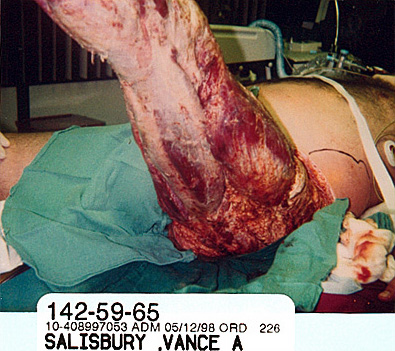
This is the post-operative photograph of my leg after the final debridement. The black line on my hip marks the perimeter of the infection, which had traveled up into my lower back. Before I was taken in, the decision was made that the infected tissue was to be removed, but Dr. Blaisdell did not expect me to live because I had lost so much tissue and the infection was spreading. However, when they began to probe, they found that the infection had miraculously disappeared and my leg was ready for skin grafts. I think this qualifies as a miracle.
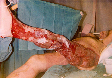
This view of my thigh from another angle shows the only area where I lost muscle, at my hip. I was fortunate — only soft tissue was affected, avoiding serious muscle loss or amputation. If you look at my hand, you can see how swollen my body was as a result of sepsis/organ failure.
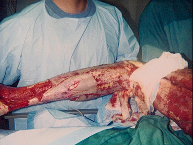
After a skin graft operation on a Tuesday, Dr. Blaisdell told me that there would be a large hole between the Achilles tendon and my ankle. It would be “flapped” or filled in by the plastic surgeon after I had been home for a month or so and gained strength. The following Saturday my cast went bad and a decision was made to take it off and clean things up a bit. When the cast was removed, they found that the flesh had covered the entire area around my ankle and was ready for skin grafts. I was awake during the procedure and heard the initial gasps when the surgeons saw the ankle, as well as another problem area on my thigh. At first, I thought it was bad news. Then, they all began saying things like “look at the ankle!” “Wow!” “It’s beautiful.” Dr. Mayer went down to the waiting area, giddy and jumping up and down. She was so excited to bring my wife the good news! Before noon, that was the worst day of my life — that afternoon was one of the most glorious, peaceful days I have ever enjoyed. I slept real sleep for the first time in the hospital that afternoon. If you look at my right leg, you will see the scarred areas where skin was “harvested.” You can also see the scars on my stomach and up my left side. I still retain the patterns on my calf and shin.
Mark, the physical therapist in the burn unit, heard the commotion and walked by my room. He came in and asked if he could work my knee while the cast was off and I said, “sure!” This is Mark holding my leg before the therapy. Note the beautiful bed of tissue God miraculously wrapped around my ankle and tendon! Mark worked my leg against his body, grasping it firmly in both hands. When he was done, he came around the side of my bed, covered in blood and gook — he looked like a butcher! Mark smiled and began snapping off his gloves and removing his gown. “Thanks, Mr. Salisbury,” he said, in his usual perky, upbeat tone,” I’ll see you later.” I looked at him and said, “You’re my hero!” My hat is off to anyone who could do what he did that day.
When my cast was removed, the surgeons were surprised and pleased to see that the graft on my thigh had “taken.” In fact, this photo shows that my grafts were doing very well and they were making good progress in reconstructing my leg. The skin retained the waffle pattern and was getting thicker.
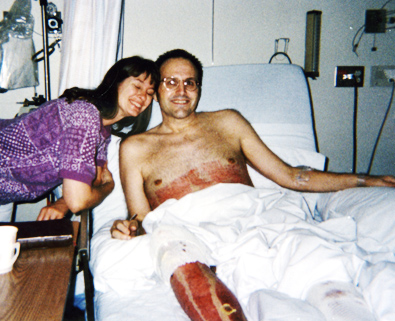
In this photo, Denise is visiting me in the trauma ward. My left leg is still in the cast and you can see the donor sites for the skin grafts on my right leg and chest. At this point, my right arm is wasting from the positioning injury to my brachial plexus and I am beginning to look like a concentration camp survivor.
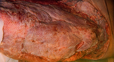
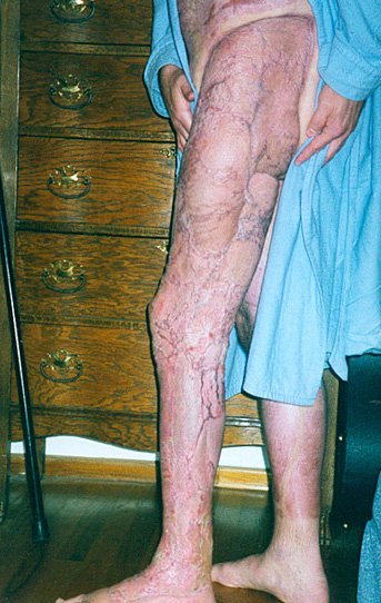
This is a photo of my finished leg. The dark lines are not veins, but the scarring around the perimeter of the individual skin grafts. The muscle has atrophied and is quite small, but my legs are back to their normal size now. You can still see the pattern of the donor sites on my right leg, where skin was harvested to graft on the affected leg. You can also see the scarring of the donor sites on my hip and stomach.

Be First to Comment